Illustrations of disease with the microscope. clinical investigations, aided by the microscope, and by chemical reagents, with microscopical observations of pathological specimens, medical and surgical, obtained in Charleston, S.C. : a contribution intended to disclose the minute history of the diseases prevailing in this latitude, and to assist future students (1861)
Porcher, Francis Peyre, 1825-1895, Billings, John S. (John Shaw), 1838-1913 · Charleston, S.C., C.S.A. : South Carolina Medical Association · leaf 104
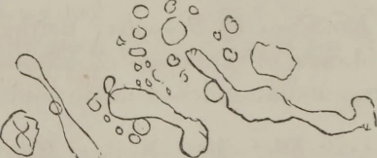
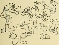
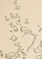
ng blacker than 1 have ever seen with nitricacid (purpurine). Examined on -Id. Still entire absence of crystals, unlessthe hsematin depicted above. Another Specimen (.nun-hud on 6th Sept. Odorpeculiar ; yellow globules,with prismatic rings—veryyellow color; also, yellowmasses and globules,… … 98 ILLUSTRATIONS OF DISEASE CaseWh.—Lawson: Recovery. Specimen examined 17th,collected 16th,[1856 ; sick five days with Yellow Fever;aet. 21. Upper surface : Vibriones small, active.
Microscopy
Internet Archive · Medical Heritage Library · Public Domain Mark 1.0. Free to use for anything.