Sthohr's histology : arranged upon an embroyological basis (1906)
Sthohr, Philipp, 1849-1911, Lewis, Frederic Thomas, 1875 · Philadelphia : P. Blakiston's Son & Co. · leaf 233
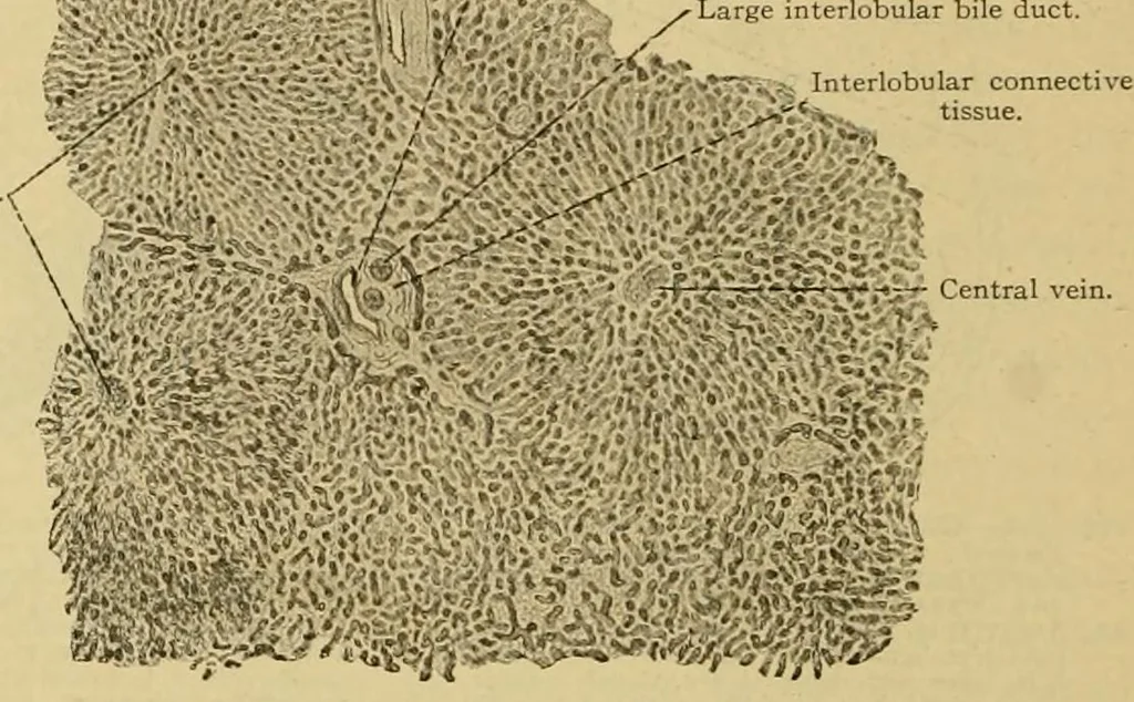
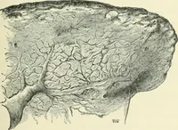
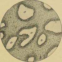
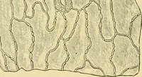
the vena cava the umbilical vein is called the ductus venosus.It follows the hepatic attachment of the lesser omentum, and there it formsafter birth, the ligament of the ductus venosus. Development of the connective tissue and hepatic artery. The historyof the liver has been described to that point… … Fig. 250.—From a Tangential Section op the Human Liver. X 40.The three central veins in cross section mark the centers of three lobules, which are not sharply separatedat the periphery from their neighbors. Below and at the right the lobules are cut obliquely andtheir boundaries are not. duct. Its…
Histology · Microscopy
Internet Archive · Public Domain Mark 1.0. Free to use for anything.